|
Early
Detection Saves Lives│中文版本│
|

|
| |
Breast
thermography has been researched for over 30 years,
and over 800 peer-reviewed breast thermography studies
exist in the index-medicus.
In this data base well over 250,000
women have been included as study participants.
The numbers of participants in many studies are
very large ranging from 37,000
to 118,000 women. Some of these studies have
followed patients up to 12 years. Breast thermography
has an average sensitivity and specificity of 90%.
|
Over 192,000 women will be diagnosed with breast
cancer in the US and 1.2 million worldwide (Source:
American Cancer Society and WHO). Breast cancer
is the top cancer among women in Hong Kong since
1994. There were nearly 2000 new patients with breast
cancer in 2001. Accordingly 1 in every 23 Hong Kong
women will have breast cancer in their life time.
The compared to western countries where 1 in every
10 women will have breast cancer, Hong Kong is lower
in incidence. However, Hong Kong is above world
average in the incidence of breast cancer. Judging
from local statistics the numbers and rates are
expected to rise steeply in the years to come. (Source:
Hong Kong Cancer Fund)
Thermography
has been around since the late 1960s and approved
by the FDA in 1982. Breast thermography is a new,
non-invasive imaging procedure that utilizes infrared
heat-sensing technology to detect metabolic changes
in the breast. Abnormalities can be detected long
before a tumor is present. It is a valuable procedure
for alerting your doctor/ physician to changes that
can indicate early stage breast disease. The benefit
of breast thermography is that it offers the opportunity
of earlier detection of breast disease than has
been possible through breast self examination, doctor
examination or mammography alone.
An abnormal thermographic
image is the single most important sign of high
risk for developing breast cancer, 10 times more
significant than a first order family history of
the disease. Studies show that this technology has
the ability to warn a woman that a cancer may be
forming up to 10 years before any other test can
detect it. This gives breast thermography not only
the ability to detect cancer at its earliest and
most treatable stage, but to also act as a biological
marker warning a woman about her own unique level
of future risk for breast cancer.
Basics of Thermal Imaging
Thermography is a non invasive test. This means
that it sends nothing into your body. In fact, there
is no contact with the body of any kind, no radiation
and the procedure is painless.
Utilizing very sophisticated infra-red cameras and
desk top computers, thermal imaging technicians
simply capture a photograph of the breasts - an
infra-red photograph (thermogram), or heat picture.
The data is stored in a computer and then can either
be printed on high resolution color printers, or
sent electronically to a physician with a similar
computer for analysis.
The physician, such as a radiologist or thermal
imaging specialist, then compares the heat patterns
in the left breast to the right breast. Any difference
in heat or any specific blood vessel patterns in
one breast that does not appear in another indicate
a physiologic abnormality. This may be pathological
(a disease) or it might indicate an anatomical variant.
When a thermogram is positive, the job of differential
diagnosis begins.
This is all that thermal imaging, or thermography
provides. A physiologic marker that some abnormality
is present in the breast. Nothing more and nothing
less. This is however, an extremely valuable and
important finding, but it has historically been
the interpretation of these findings that has been
the problem, and is now the subject of the "responsible
second look".
|
| Reasons
to Choose Breast Thermography? |
Early detection
Non invasive
No radiation
Painless
No contact with the body
F.D.A approved*
Remarks: FLIR A-series telethermographic camera
is applied in the Breast Thermography, and it
is designated by the FDA under Section 510(k),
for the following indications of use:
- The FLIR devices are intended for uses as an
adjunct to other clinical diagnosis, quantifying,
and screening of differences of skin surface temperature
changes.
- It can visualize. Document temperature patterns
and changes.
- The environments of use are: hospitals, sub-acute,
public areas (i.e. airports), etc.
With the incidence of breast cancer steadily rising
in women under 40, an effort to provide some form
of accurate screening test is needed in this age
group. Very early detection is especially important
since breast cancers in younger women are commonly
more aggressive resulting in lower survival rates.
Current screening procedures have proven to be
inaccurate in women in this age group due to breast
tissue density and other factors. These issues,
however, do not affect thermography. With this
technology, women under 40 now have a safe and
objective screening method that they can add to
their regular breast health check ups.
Currently, no single screening procedure can detect
100% of all breast cancers. Thermography is designed
to be used with mammography and not as a replacement.
Studies show that when thermography is added to
a woman's regular breast health check ups (physical
examination + thermography + mammography), 95%
of all early stage cancers will be detected.
Competition Paradox with Mammography
Scientists and health care researchers have been
looking for many decades at tools that can identify
breast cancer reliably and quickly. It takes years
for a tumor to grow, and the earliest possible
indication of abnormality is needed to allow for
the earliest possible treatment and intervention.
Thermography is a test of PHYSIOLOGY. It does
not look at anatomy or structure, and it only
reads the infra-red heat radiating from the surface
of the body.
Mammography, on the other hand, is a test of ANATOMY.
It looks at structure. When a tumor has grown
to a size that is large enough, and dense enough
to block an x-ray beam, it produces an image on
the x-ray or mammographic plate, that can be detected
by a trained radiologist. A fine needle biopsy
is then generally performed to identify the type
of tissue in the mass, to determine if atypical
or cancerous cells are present.
We now come to an important point. Neither thermography
nor mammography can diagnose breast cancer. They
are both diagnostic tests which reveal different
aspects of the disease process and allow for further
exploration. |
|
| Who
Should Have This Screening? |
All women can benefit from breast
thermography screening. However, it is especially
appropriate for younger women (30 - 50) whose denser
breast tissue makes it more difficult for mammography
to be effective. Also for women of all ages who,
for many reasons, are unable to undergo routine
mammography. This test can provide a 'clinical marker'
to the doctor or mammographer that a specific area
of the breast needs particularly close examination.
It takes years for a tumor to grow thus the earliest
possible indication of abnormality is needed to
allow for the earliest possible treatment and intervention.
Thermography's role in monitoring breast health
is to help in early detection and monitoring of
abnormal physiology.
The American Cancer Society says women are at risk
for breast cancer if they:
|
|
Began menstruating before
age 12 |
|
Have not borne children |
|
Bore their first child after age 30 |
|
Stopped menstruating after age 50 |
|
Have a personal history or family history of breast
cance |
|
Have a personal history of radiation exposure
to the chest |
|
Are currently taking or have recently taken hormone
replacement therapy (HRT) for longer than five years |
|
Are obese |
| |
| Guideline
for Breast Thermography Screening: |
| Age 20 - baseline
thermogram
Ages 20 to 30 - every three years
Ages 30 and over - every year
Statistics indicate that 1/3 of all breast cancers
occur between the ages of 20 and 44 years of age.
These guidelines, therefore, include careful breast
monitoring during these years. With the addition
of thermography, interval cancers (cancers which
show up between mammograms) are thought to be
detected much sooner. |
|
| What
If Breast Thermogram Results Are Positive? |
All medical screening tests, including
thermography and mammography, are just that - screening
tests.
Any positive screening test requires further evaluation.
Breast thermograms receive one of five ratings that
range from TH1 (no detectable thermal abnormalities)
to TH5 (detection of thermal abnormalities correlating
with very significant risk for breast cancer). Early
thermal abnormalities may result in a recommendation
to repeat thermography for comparison in 60-120
days. Depending on the thermology rating and clinical
findings, a referral may be made for targeted ultrasound
or to a breast specialist for possible biopsy.
Physicians trained in holistic medicine may also
recommend nutritional, metabolic, environmental,
or lifestyle interventions to address early thermal
abnormalities.
Thermography is a screening tool for breast cancer
that is best utilized with regular breast self-exams
(BSE) and annual clinical breast exams (CBE) by
a healthcare professional. |
|
|
|
|
|
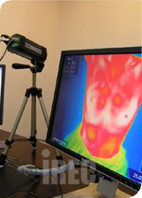
Infrared Thermography Station
|
|
|
|
|
|
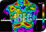
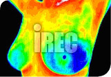
A woman stopped nurturing
for a year
|
|
|
|
|
|
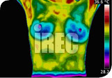
Breast Implant
(Souce: Image of Health)
|
|
|
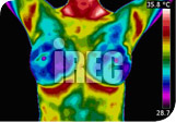
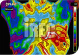
TH-5 Abnormal, Turn or Confirmed
(Source: breastthermography.com)
|
|
|